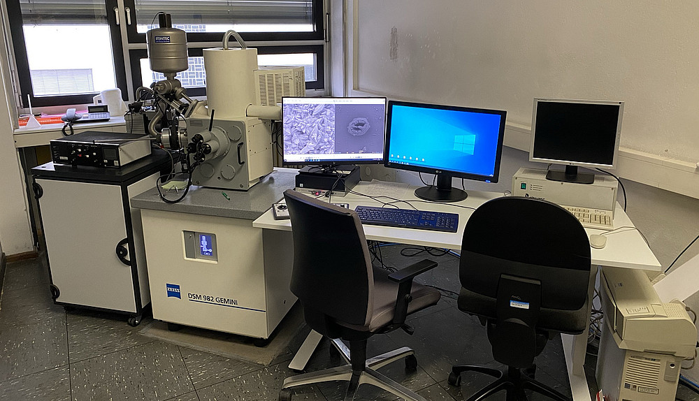
The Institute of Earth Sciences has a modernised field emission scanning electron microscope (FE-SEM) from Zeiss, which allows resolution in the sub-nanometre range (up to 300,000x magnification depending on the nature of the sample).
The FE-SEM can be used to visualise the smallest objects and determine their composition. In this way, details of the origin and evolution of life can be researched, but also new mineral building materials such as cement can be developed. By determining the chemical elements in microscopic minerals, processes of the dynamic earth can also be analysed.
Two SE detectors: (InLens and chamber detector)
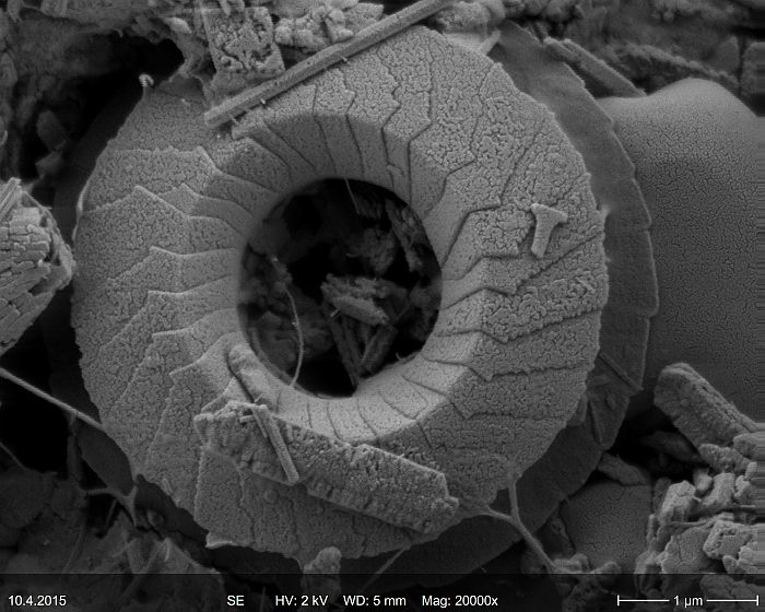
Both secondary electron detectors are used for surface imaging in the micron to sub-nanometer range. Images can be captured in a short time and with minimal preparative effort.
BSE detector
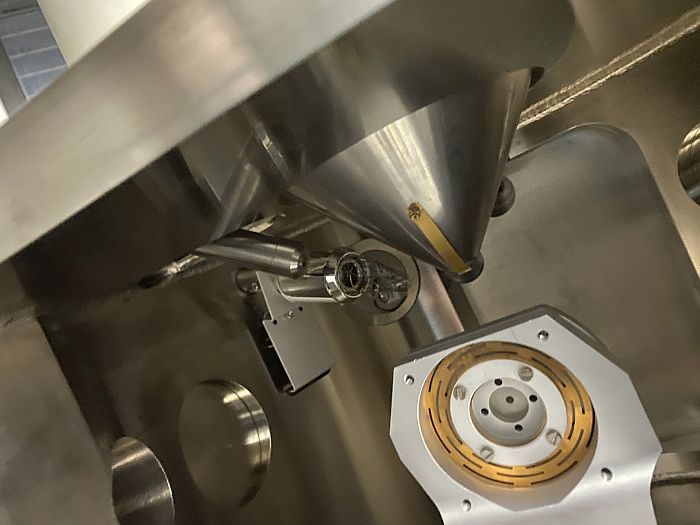
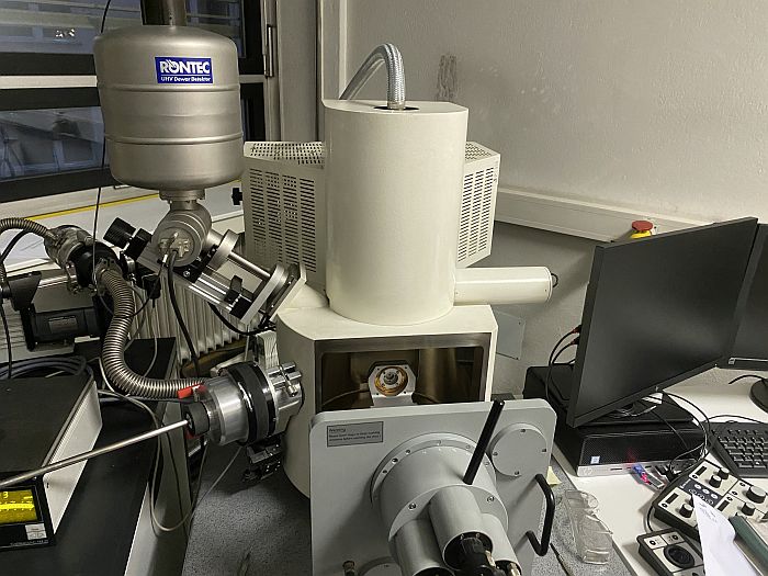
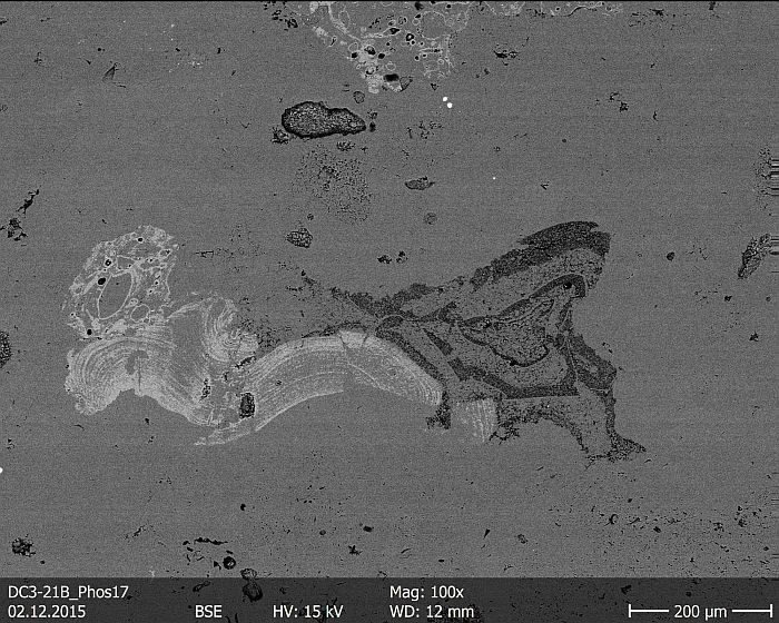
The externally mounted backscattered electron (BSE) detector is used to map the atomic number contrast of materials within a sample. This method allows different material phases in a sample to be distinguished quickly and easily on the basis of their chemical/elemental composition. Materials that are composed of relatively heavier elements appear brighter.
Energy dispersive X-ray microanalysis system
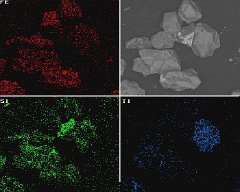
The SEM is also equipped with an energy dispersive X-ray microanalysis system, EDX for short. With this analytical add-on, element distribution images can be generated in the samples to be analyzed.
The measuring principle is as follows: When the electron beam used to generate the image interacts with the sample, X-rays characteristic of the elements contained in the sample are produced, among other things. The detector receives this radiation and displays the signal either as an energy spectrum or as a flat image with different colors for individual elements. This allows the chemical composition of examined samples to be determined quickly.
