A JXA-8530F field emission microprobe from JEOL has been in use at the Department of Earth Sciences since 2018. This can be used to generate high-resolution images and carry out non-destructive chemical analyses in the major, minor and trace element range.
How it works:
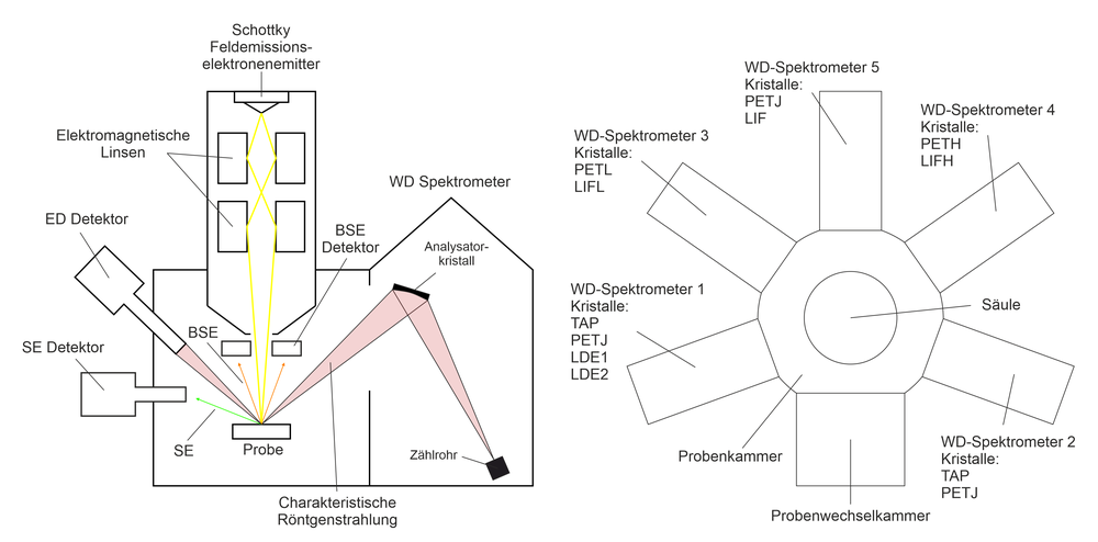
A Schottky field emission electron emitter and several electromagnetic lenses are used to generate a fine electron beam (up to <1 µm in diameter) (Figure 1). This beam hits the sample, causing interactions between the electrons in the electron beam and the atoms in the sample. These interactions are used to analyze chemical compositions but also for imaging.

Chemical analyses:
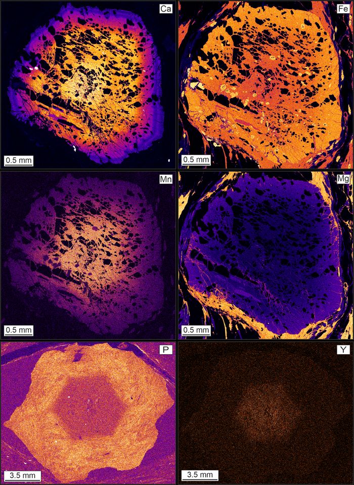
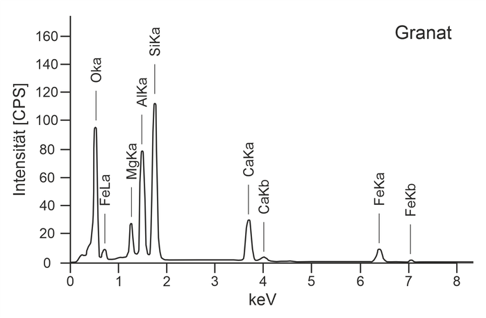
The interaction between the electron beam and the sample produces, among other things, characteristic X-rays that have element-specific wavelengths and energies. Using wavelength dispersive (WD) X-ray spectroscopy or energy dispersive (ED) X-ray spectroscopy, the intensities of these X-rays are measured in order to determine the chemical composition of the sample in percent by weight. The field emission microprobe at the Department of Earth Sciences is equipped with five WD spectrometers. This allows up to five elements with an atomic number of 5 (boron) to 92 (uranium) to be measured simultaneously during a point analysis or mapping (element distribution image; Figure 2). Furthermore, an ED detector is also built in, with which the entire X-ray spectrum of a mineral can be generated in a very short time (Figure 3).


Imaging
During the interaction between the electron beam and the sample, secondary electrons (SE) and backscattered electrons (BSE) are also generated, which are used for imaging. The JXA-8530F field emission microprobe has both a BSE and an SE detector, which allows an image to be generated when the sample surface is scanned with an electron beam. SE images are mainly used to examine surfaces, while BSE images can be used to draw conclusions about the chemical composition (e.g. zoning) (the brighter the image, the higher the average atomic number of the mineral; Figure 4). Furthermore, this field emission microprobe is also equipped with a cathodoluminescence (Cl) detector and an optical microscope (reflected light microscope).
Application
At the Department of Earth Sciences, the field emission microprobe is mainly used for routine analyses of major and minor elements of rock-forming minerals (garnet, feldspar, mica, amphibole, pyroxene, olivine, chlorite, staurolite, cordierite, etc.). The minerals are analyzed both quantitatively and qualitatively with a sample current of 10 nA and a beam diameter of 1-5 µm (Figure 2). For quantitative measurements, the determined intensities of the elements are converted into weight percentages using reference materials (standards). Some of the reference materials used are "Smithsonian Microbeam Standards". The Institute of Earth Sciences would like to take this opportunity to thank the Smithsonian for providing these standards.
Another field of application of the field emission microprobe is the analysis of trace elements and rare earth elements (La to Lu). With a sample current of 50 to 150 nA, e.g. Y and P contents in garnet (Figure 2), Zr contents in rutile and titanite (geothermometer) or rare earth element contents in monazite, allanite, florencite, as well as other "exotic" minerals such as Y-gaddolinite are measured.
The field emission microprobe is also used to carry out chemical dating of monazite. Monazites are identified in thin section using BSE images (Figure 4) and then measured in situ with a sample current of 150 nA and a beam diameter of 3 to 5 µm. The small beam diameter also makes it possible to date fine zoning in the monazite. In particular, the exact content of Th, U and Pb is required for accurate dating, i.e. U and Pb are measured in total for 400 seconds (200 s at the peak and 100 s at the background). Th is analyzed with shorter counting times (20 s at the peak and 10 s at the background), but on two different spectrometers in order to obtain the most accurate result possible. The age is determined from the chemical analyses based on the method of Suzuki et al. (1991).
Publications
- Stumpf, S., Skrzypek, E., Stüwe, K. (2024). Dating prograde metamorphism: U-Pb geochronology of allanite and REE-richepidote in the Eastern Alps. Contribution to Mineralogy and Petrology, in press.
- Sorger, D., Hauzenberger, C. A., Finger, F., Linner, M., Skrzypek, E., & Schorn, S. (2024). Formation oflow-pressurereaction textures during near-isothermalexhumation of hot orogenic crust (Bohemian Massif, Austria). Journal of Metamorphic Geology, 42(1), 3-34. doi:10.1111/jmg.12744
- Schorn, S., Rogowitz, A., & Hauzenberger, C. A. (2023). Partial melting of amphibole-clinozoisite eclogite at the pressure maximum (eclogite type locality, Eastern Alps, Austria). European Journal of Mineralogy, 35(5), 715-735. doi:10.5194/ejm-35-715-2023
- He, D., Dong, Y., Hauzenberger, C. A., Sun, S., Neubauer, F., Zhou, B., Yue, Y., Hui, B., Ren, X., & Chong, F. (2022). Neoproterozoic HP granulite and its tectonic implication for the East Kunlun Orogen, northern Tibetan Plateau. Precambrian Research, 378, 106778. doi:10.1016/j.precamres.2022.106778
- Nong, A. T. Q., Hauzenberger, C. A., Gallhofer, D., Booth, J., Skrzypek, E., & Dinh, S. Q. (2022). Early Mesozoic granitoids in southern Vietnam and Cambodia: A continuation of the Eastern Province granitoid belt of Thailand. Journal of Asian Earth Sciences, 224, 105025. doi:10.1016/j.jseaes.2021.105025
References
Suzuki, K., Adachi, M., & Tanaka, T. (1991). Middle Precambrian provenance of Jurassic sandstone in the Mino Terrane, central Japan: Th-U-total Pb evidence from an electron microprobe monazite study. Sedimentary Geology, 75(1-2), 141-147.
