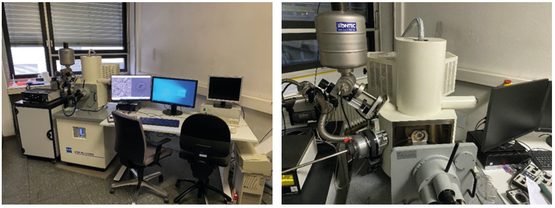
Field Emission Scanning Electron Microscope
The Gemini DSM 982
The of Institute of Earth Sciences runs a refurbished Field Emission Scanning Electron Microscope (FE-SEM). The FE-SEM provides easy and fast imaging capabilities with resolutions down the sub-nanometer scale (up to 300.000x magnification depending on sample conditions).
A field emission cathode is used in this device to generate the electron beam. There are four detector systems available for image generation:
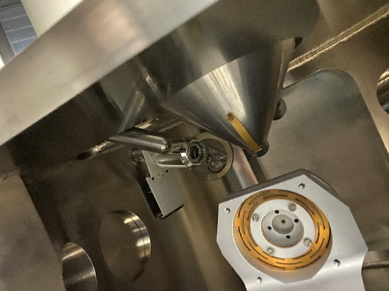
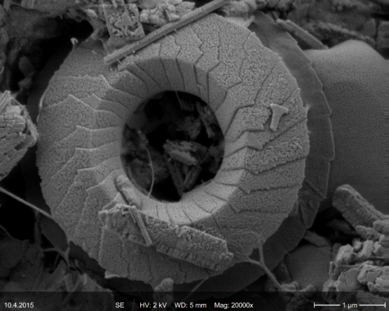
Two SE-Detectors: (InLens- und chamber-detector)
Both secondary electron (SE) detectors are used for surface imaging on micron to sub-nanometer resolutions. Images can be taken in a short time and with only minimal preparative effort.
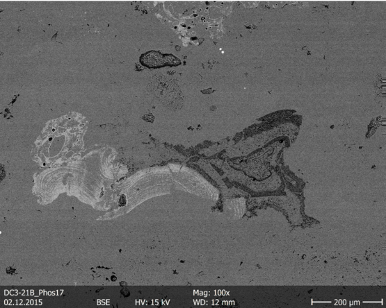
BSE-Detector
The externally mounted backscattered electron detector (BSE detector) is used to map the atomic number contrast of materials within a sample. This method allows different material phases in a sample to be quickly and easily differentiated based on their chemical composition. Materials that contain relatively heavier elements appear brighter in the image.
CL-Detector (ab Sommer 2021):
In the summer of 2021, the institute's FE-SEM will be expanded to include a CL detector. Further information will follow.
The Energy Dispersive X-ray Microanalysis System
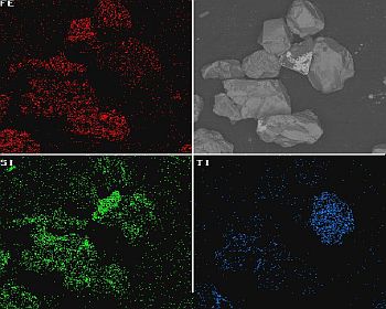
The FE-SEM is also equipped with an energy-dispersive X-ray microanalysis system for short EDX. With this analytical add-on, element distribution images can be generated in the samples to be analyzed.
The measuring principle is as follows: When the electron beam used to generate images interacts with the sample, an X-ray radiation that is characteristic of the elements contained in the sample is generated. The detector receives this radiation and displays the signal either as an energy spectrum or as a flat image with different colors for individual users. This also enables the chemical composition of the examined samples to be quickly determined.